Read more news
Filter by
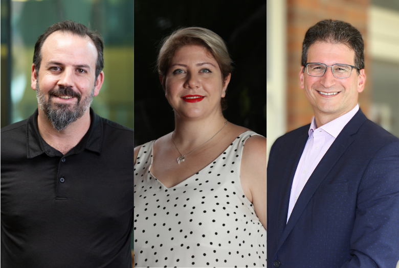
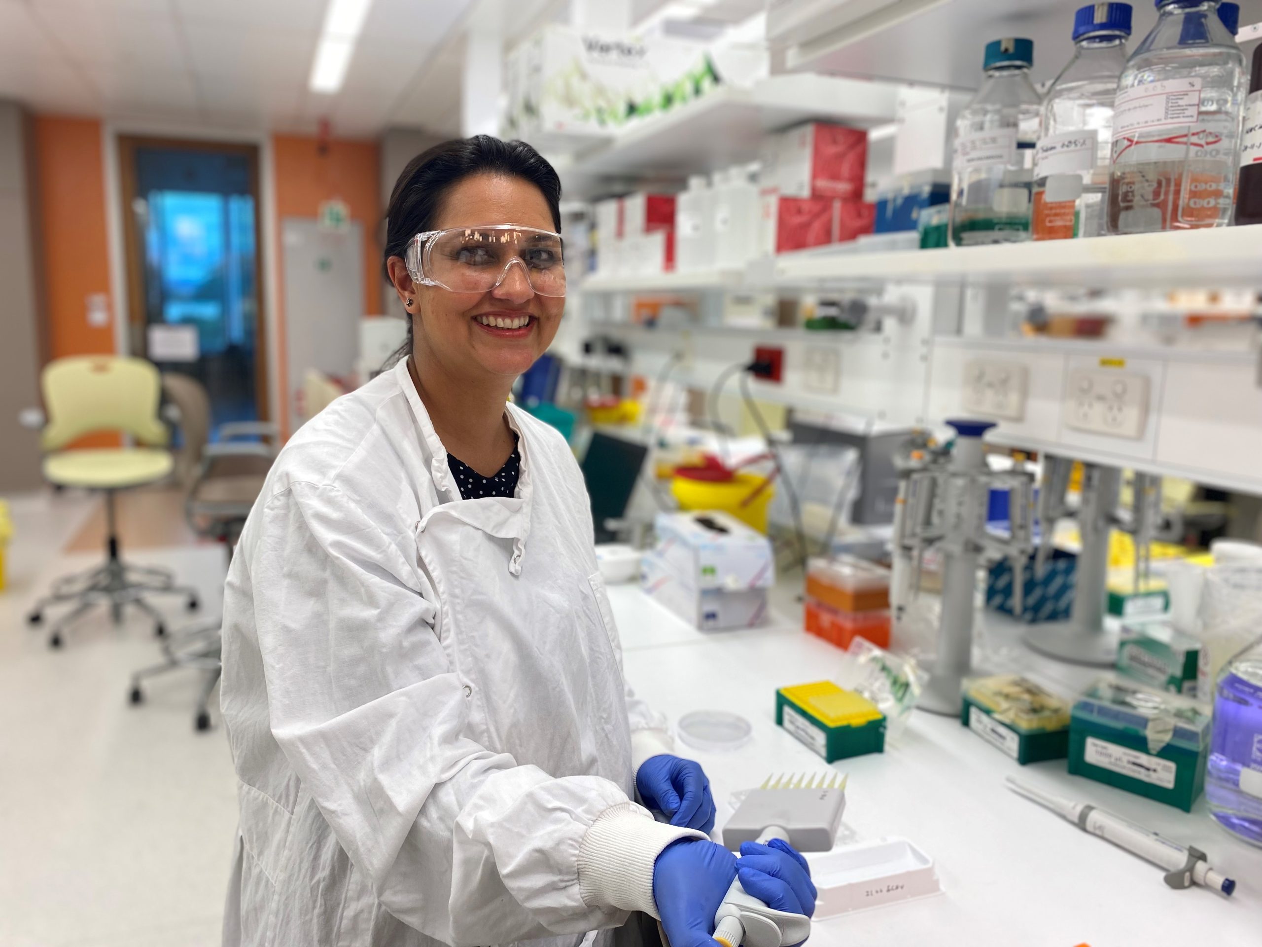
In a bone tumour microenvironment model developed by QUT biomedical scientists led by TRI-based Dr Nathalie Bock, they found anti-androgen treatment helped prostate cancer cells to adapt and grow.
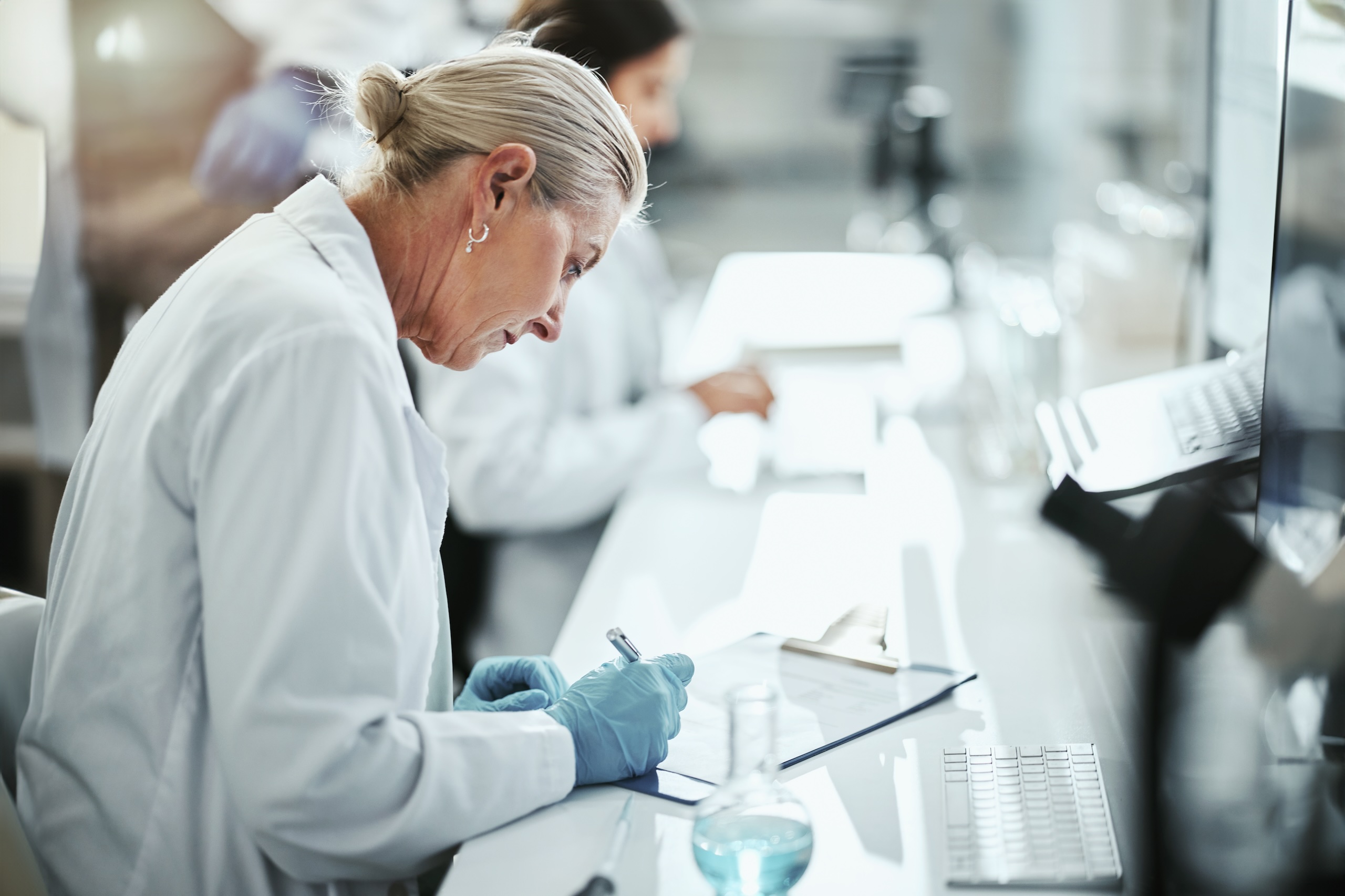
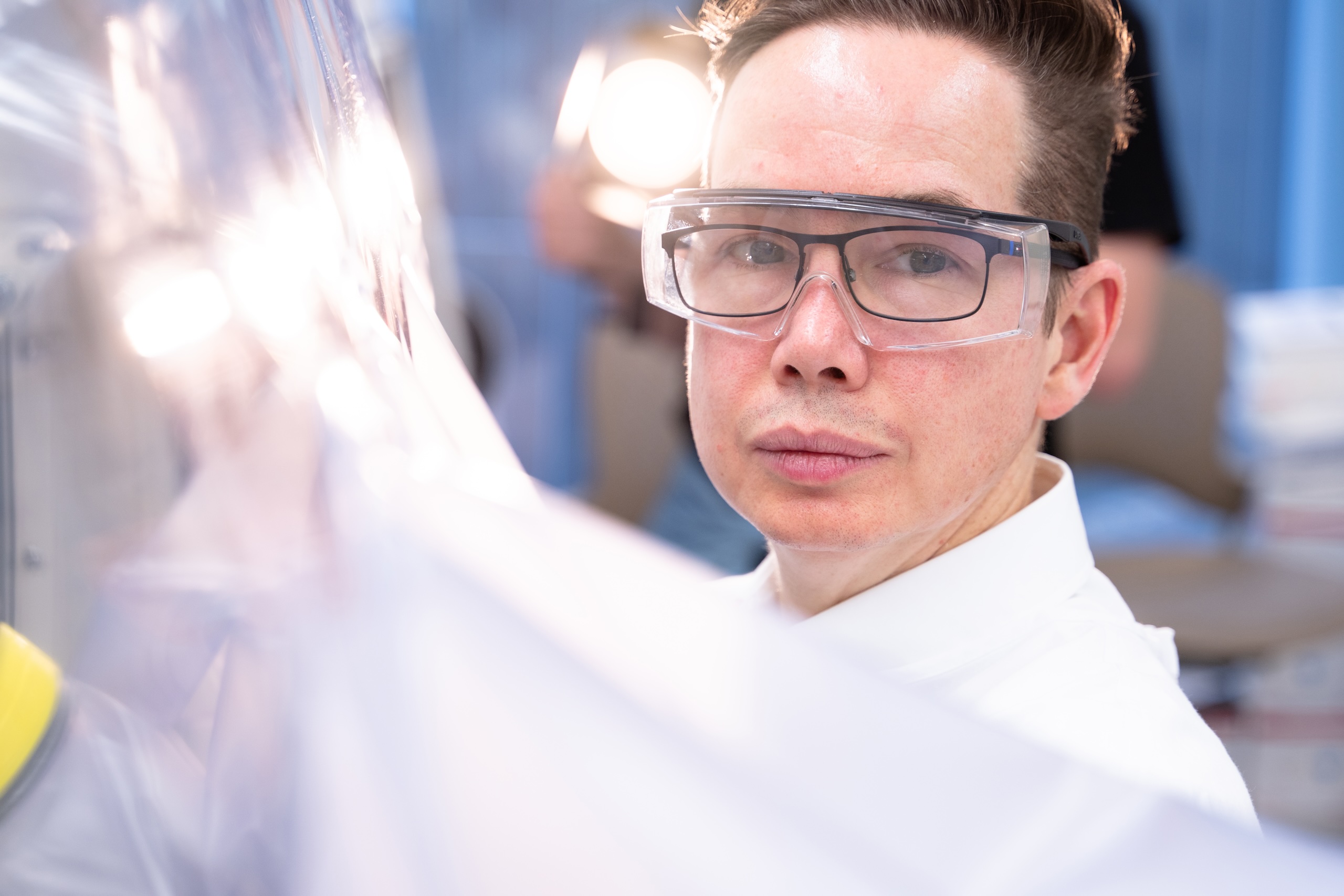
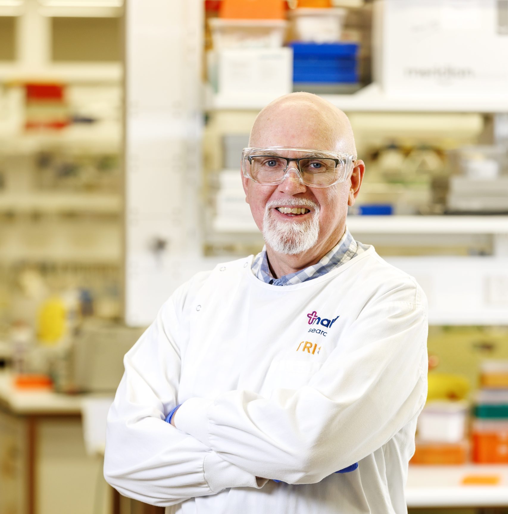
Dr Bock, under the mentorship of Distinguished Professor Dietmar Hutmacher, from QUT’s Centre for Biomedical Technologies, focused her research on bone metastases from breast and prostate cancers.
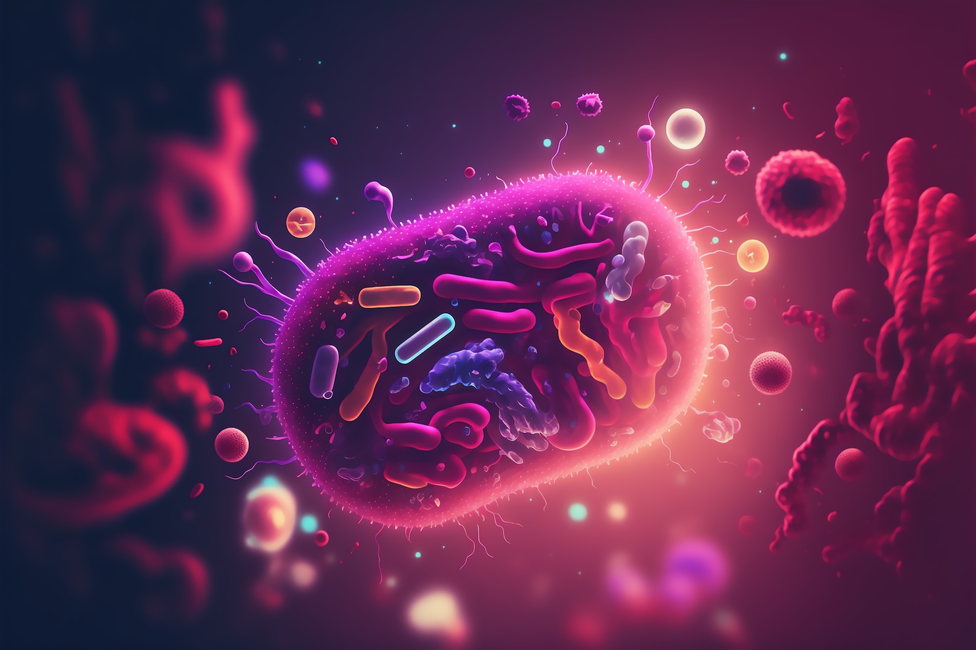
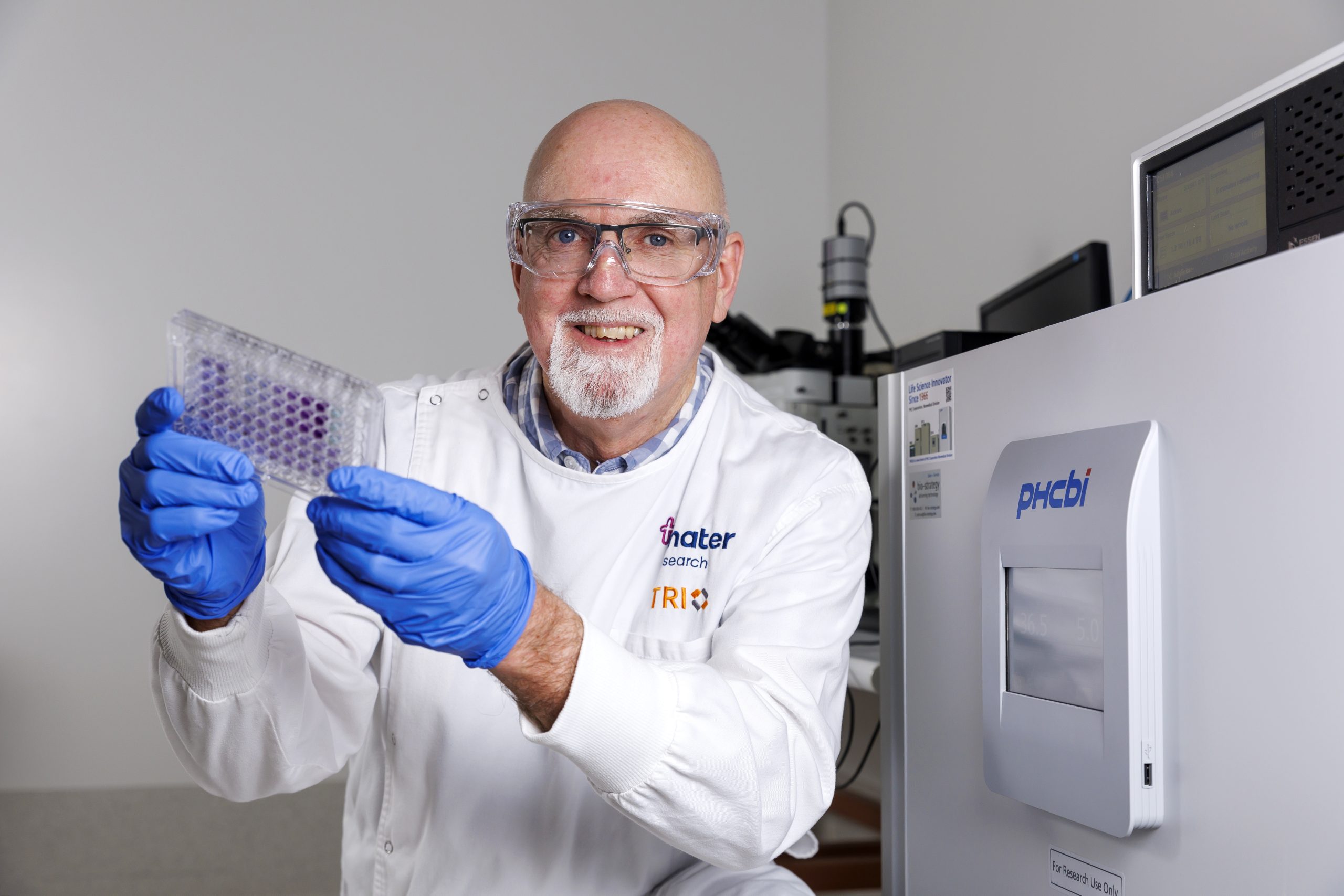
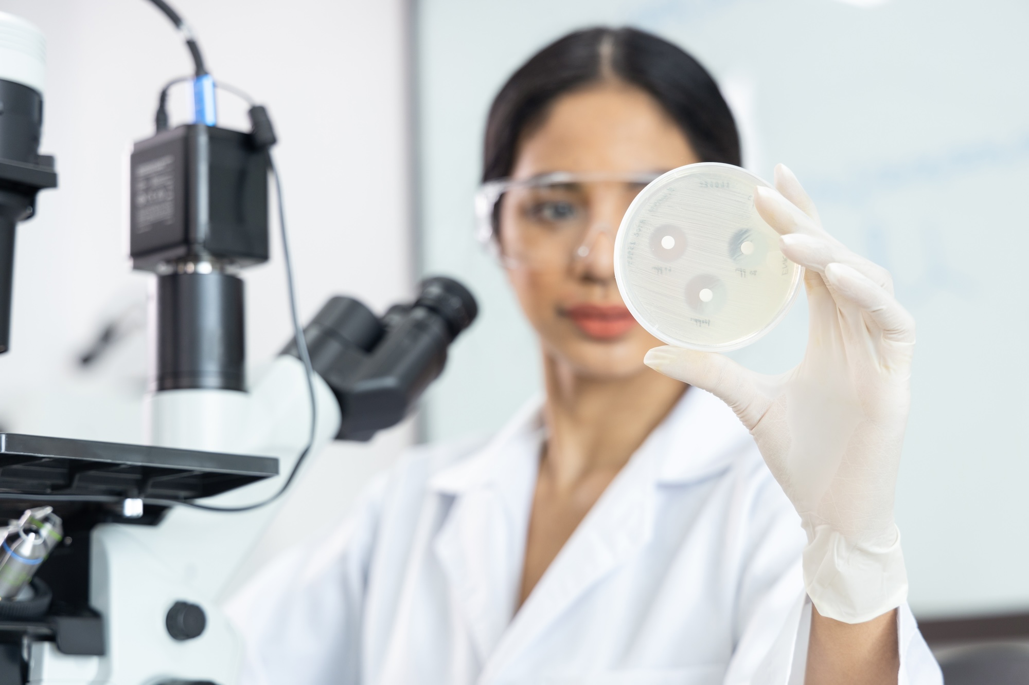
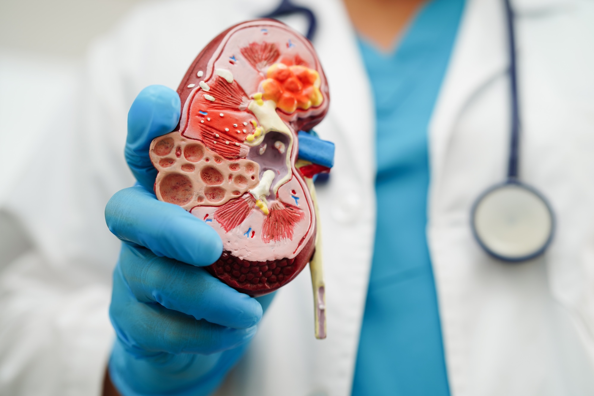
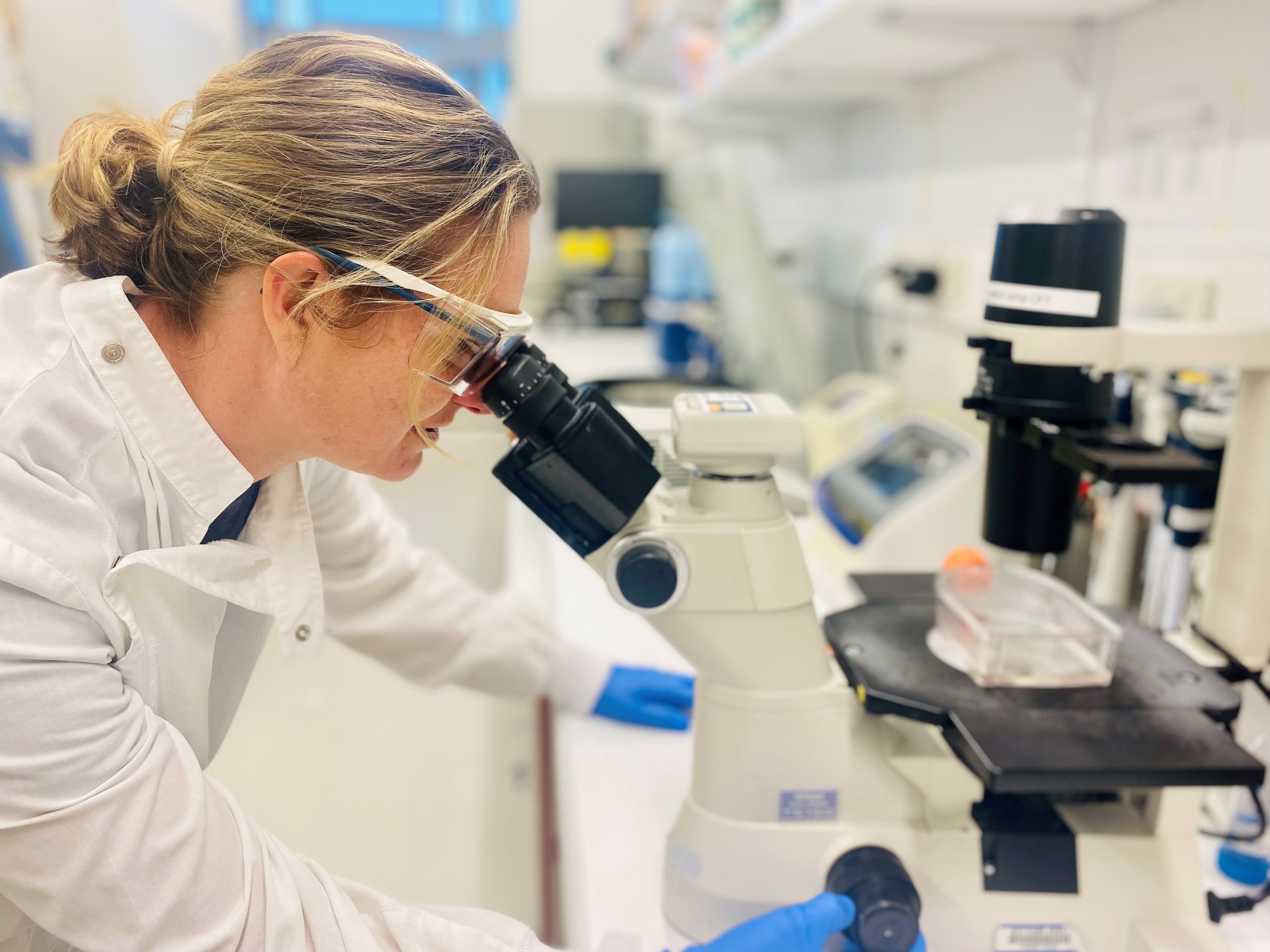
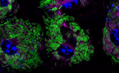
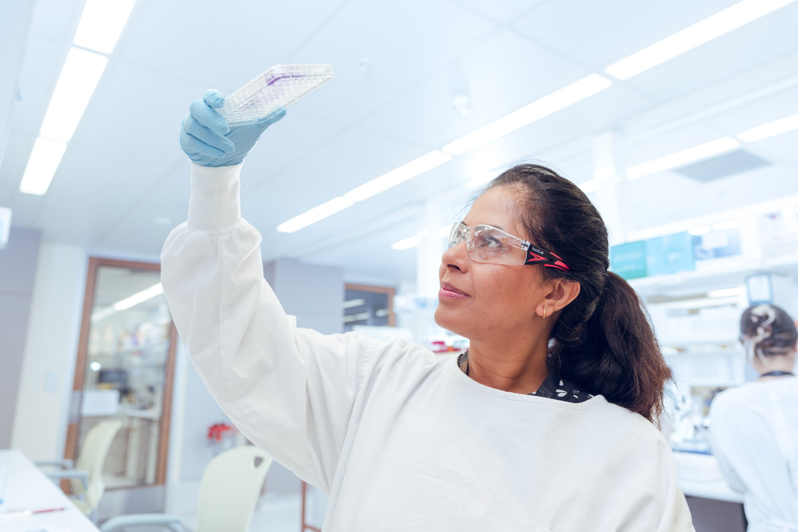
Through her work, Dr Bock developed 3D miniature bone-like tissue models in which 3D printed biomimetic scaffolds are seeded with patient-derived bone cells and tumour cells to be used as clinical and preclinical drug testing tools.
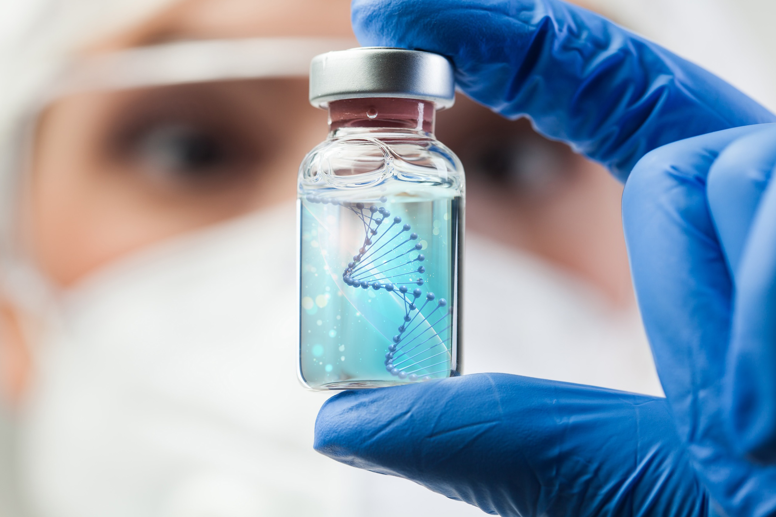
The research team investigated their hypothesis that traditional anti-androgen therapy had limited effect in the microenvironment of prostate cancer bone tumours. The team’s findings are published in Science Advances.
“We wanted to see if the therapy could be a contributor of cancer cells’ adaptive responses that fuelled bone metastasis,” Professor Hutmacher said.
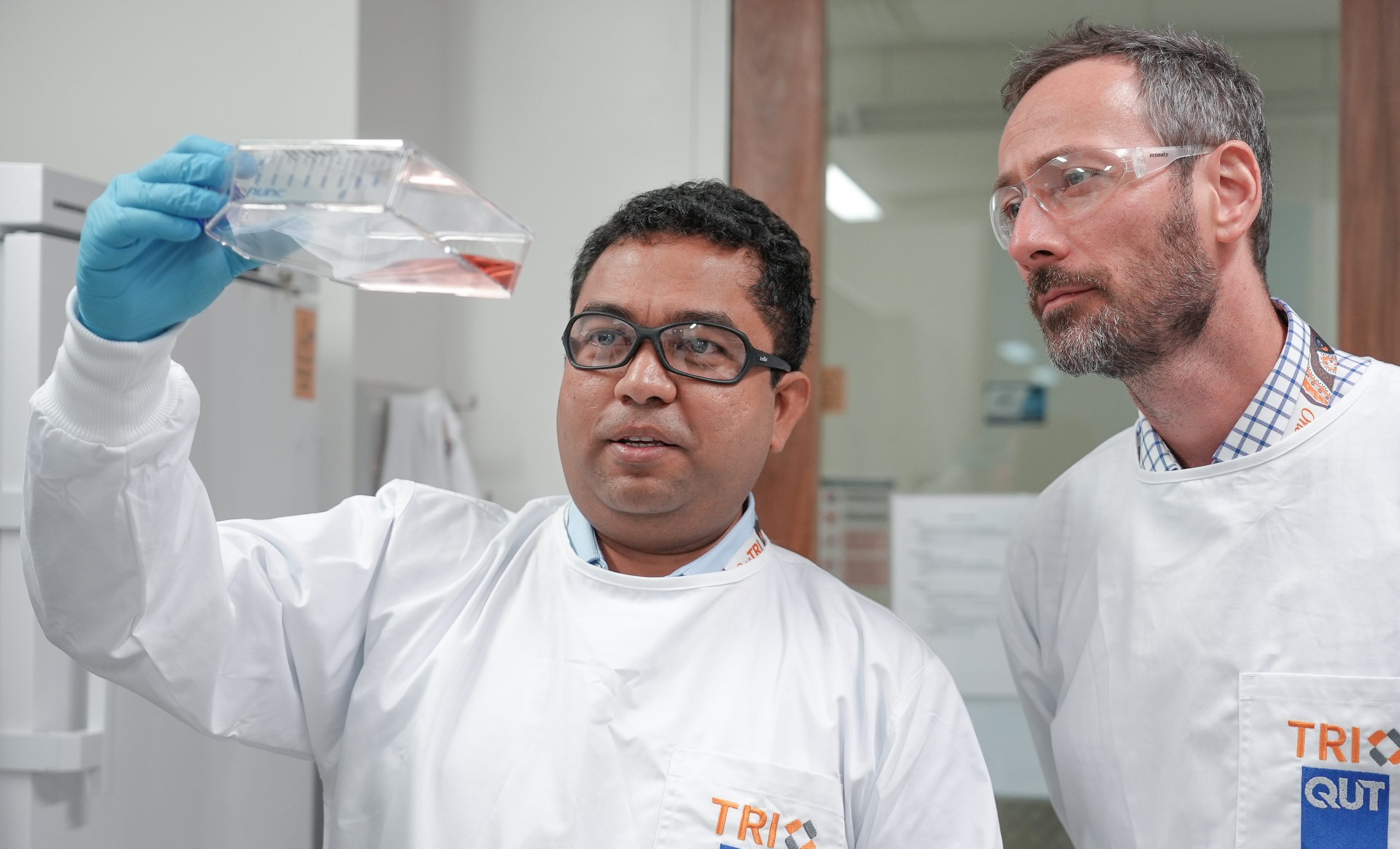
“We developed an all-human, microtissue-engineered model of metastatic tissue using human bone-forming cells, prostate cancer cells and 3D printing.”
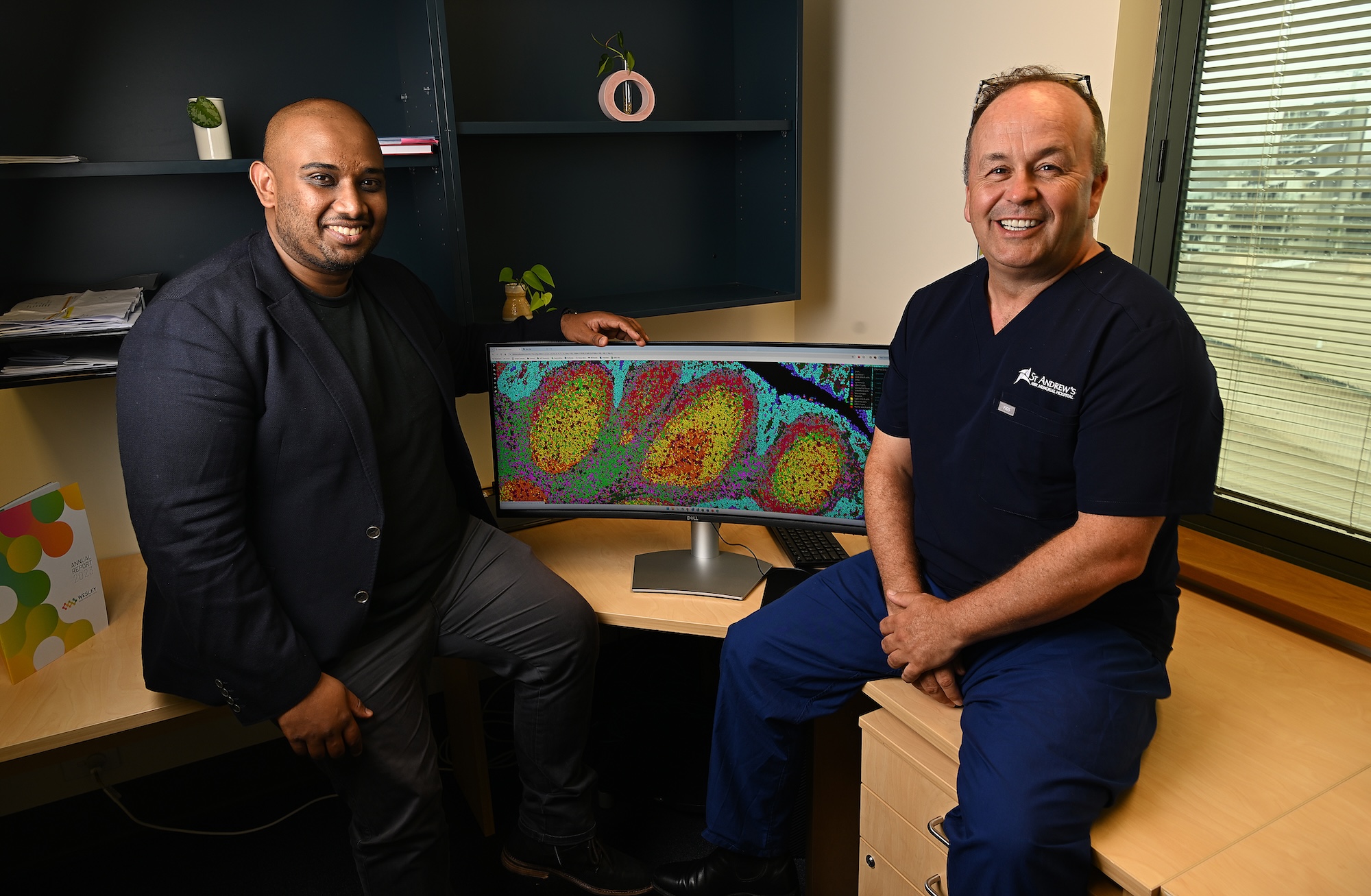
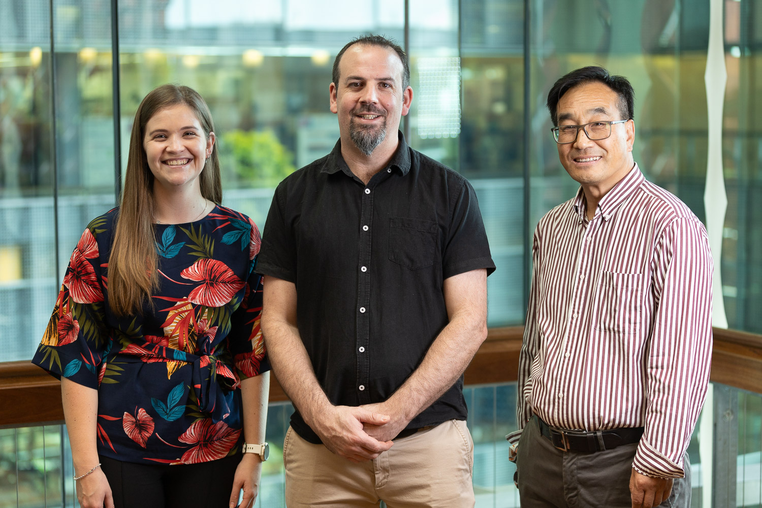
Cancer biologist Distinguished Professor Judith Clements, who is also based at TRI, said the team bioengineered the microenvironment of a bone tumour to assess the effects of two clinically routinely used anti-androgen therapies – enzalutamide and bicalutamide – on the tumour cells.
“We found that the interactions between the cancer cells, the bone and the anti-androgens significantly impacted the progress of cancer in the mineralised microenvironment of bone tumours,” Professor Clements said.
“This means that the efficacy of these therapies is compromised in the presence of the bone microenvironment.”
Professor Hutmacher said an important outcome of the study was the need to upscale the bone tumour microenvironment model platform and make it available to other research groups.
“This would enable the prostate cancer research community to develop therapies for a more effective treatment of advanced prostate cancer.”
In future, Dr Bock will use her model with patient-derived cells from patients undergoing prostatectomy, so that it could be used as a personalised preclinical diagnostic and drug testing tool.
“By screening existing and novel drugs using the bone tumour model in the laboratory, doctors will be able to treat individual patients with an anti-cancer therapy that can best suits their clinical need.”
“This has the potential to considerably improve the quality of life of patients, because patients will not have to trial a succession of drugs, each of which carries the potential of severe side-effects, and which may not work for them.”
This research was supported by the National Health and Medical Research Council of Australia, Australian Research Council and the Prostate Cancer Foundation of Australia.
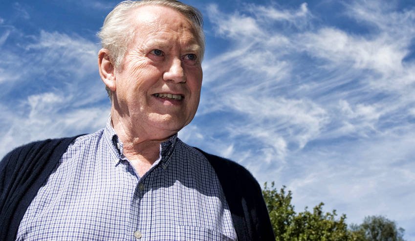
Prostate Cancer Foundation of Australia CEO Professor Jeff Dunn AO said the findings were significant.
“This is an important discovery that will help us to better target treatments for men with different types of prostate cancer,” he said.
“The findings also demonstrate the importance of ongoing research to improve our understanding of how different treatments impact disease progression and spread.
“This is Australian research excellence at its finest.”
The published paper:
Nathalie Bock et al. In vitro engineering of a bone metastases model allows for study of the effects of antiandrogen therapies in advanced prostate cancer. Sci. Adv. 7, eabg2564 (2021).DOI: 10.1126/sciadv.abg2564