Case Studies
Transforming skin cancer diagnosis in Australia
Skin cancer is a significant public health issue globally, but none more so than in Australia, where a person is more likely to die from melanoma than in a road traffic accident.
The University of Queensland (UQ) Dermatology Research Centre, based at TRI, is on the cusp of changing how Australia screens and diagnoses people for skin cancer by combining the latest in research and world-class technologies.
Key points
- VECTRA is a whole-body imaging system that creates a 3D avatar capturing a person’s skin surface, which can be used for skin cancer examination.
- The UQ Dermatology Research Centre brought the first VECTRA machine to Australia and pioneered the first clinical trial at TRI’s Clinical Research Facility.
- The recently established Australian Cancer Research Foundation Australian Centre of Excellence in Melanoma Imaging and Diagnosis is deploying 15 VECTRA systems across Queensland, New South Wales and Victoria.
- The UQ Dermatology Research Centre has also developed and licensed a minimally invasive microbiopsy device.
Australia has the highest rate of skin cancer in the world.
Squamous and basal cell carcinomas are the most common form of skin cancer and they typically develop in areas of the skin exposed to the sun, such as the face, neck and arms. While these types of skin cancer are generally less aggressive than melanoma, they still require prompt treatment to prevent complications.
Melanoma, the deadliest form of skin cancer, develops in the cells responsible for producing pigment in the skin. Once melanoma develops, it can spread to other parts of the body to form new tumours in distant organs, such as the lungs, liver, brain or bones. Once melanoma has spread it becomes much more difficult to treat and has a much poorer prognosis, so early detection is crucial.
While there has been extensive research into the best ways to identify and screen high-risk people, the current skin cancer screening landscape is fragmented and lacking a consistent approach.
Traditional methods of skin cancer detection rely on visual examination, which can be subjective and may not always capture subtle changes in moles that could indicate melanoma development.
Additionally, Australia’s vast geographical spread presents further obstacles for healthcare. Melanoma patients living in rural regions face difficulties in accessing specialist care, with a reported 20 per cent higher melanoma-related mortality rate compared to urban areas.
How an Australian first, led to the world’s largest trial of its kind
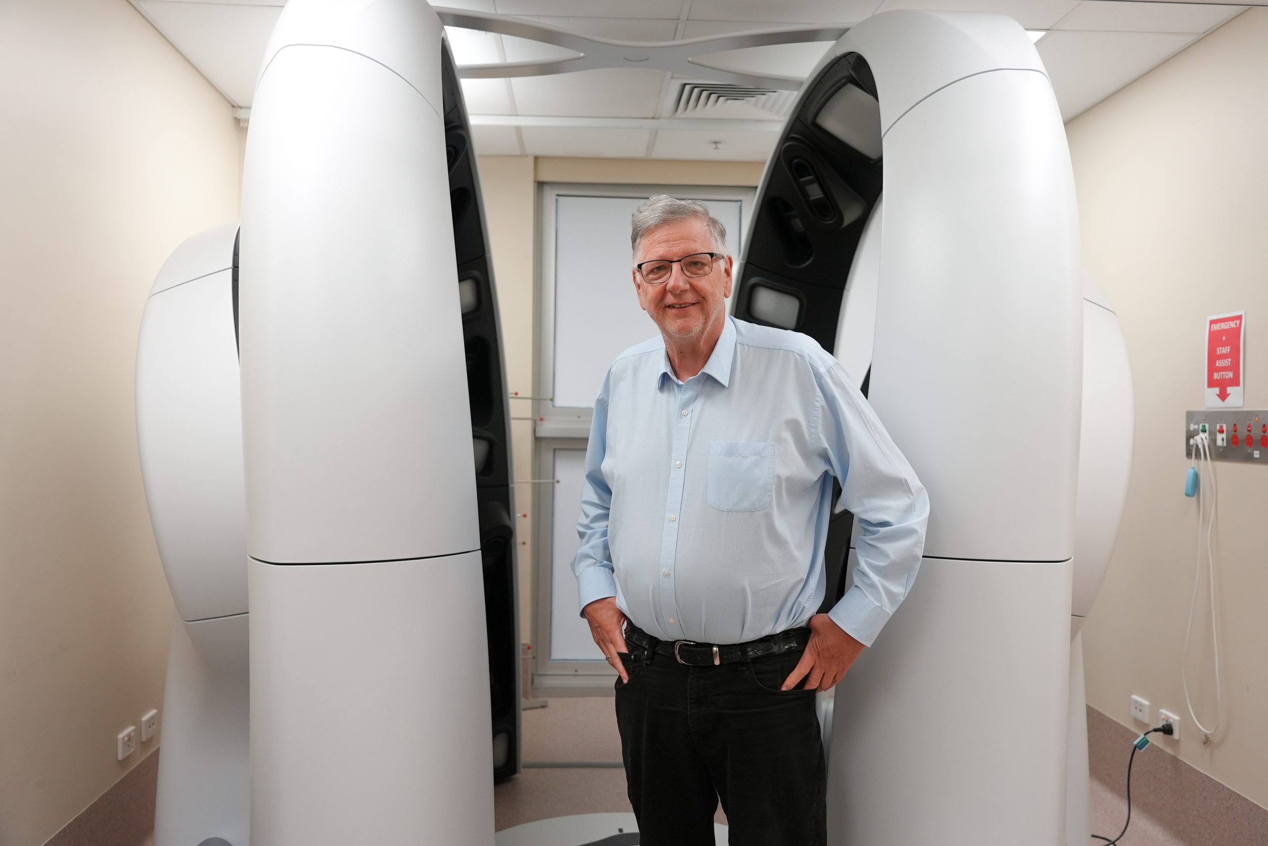
VECTRA is a whole-body imaging system that uses 92 cameras to simultaneously capture, in seconds, a 3D body avatar of a person to visualise their entire skin surface in high detail. It excludes the soles of the feet, scalp and areas covered by clothing.
The images are securely stored and accessible to clinicians for assessment, greatly enhancing the ability to track and identify changes to skin spots and moles over time.
Australia’s first prototype VECTRA imaging system was funded by the Princess Alexandra Hospital Private Practice Trust Fund and installed at TRI’s Clinical Research Facility in 2015. The system was used for several clinical research projects – and after ongoing R&D with the vendor, it was replaced with Australia’s first commercial version of the VECTRA 3D imaging system in 2017.
Following a $10 million grant awarded by the Australian Cancer Research Foundation (ACRF) in 2018, the Australian Centre of Excellence in Melanoma Imaging and Diagnosis (ACRF ACEMID) was launched at TRI in 2021. The ongoing project involved the establishment of 15 VECTRA 3D imaging systems across metropolitan and regional locations in Queensland, New South Wales and Victoria – all supported by a telemedicine research network which connects the regional and rural sites with dermatology specialists at the metropolitan hub.
UQ’s Professor H. Peter Soyer, who is Director of the Dermatology Research Centre and the Principal Investigator for the ACRF ACEMID project, said his team’s goal was to see a world free of melanoma.
“Australians are diagnosed with melanoma at 12 times the global average, and it is the most common cancer in people aged 15 to 40,” Professor Soyer says.
“Early diagnosis is critical. People with Stage 1 melanoma have a 98 per cent chance of surviving beyond five years.
“Through technologies such as telehealth, and AI, we can help make specialists more accessible and hopefully break down some of the barriers preventing people from being checked.”
Using the state-of-the-art research infrastructure at TRI, the Dermatology Research Centre team is focused on developing and validating several technologies in the fight against skin cancer.
A better way to biopsy
Another of the team’s success stories is the invention of a skin microbiopsy device that is less invasive than current ones on the market.
Skin biopsies are a standard procedure for diagnosing suspected skin cancers and inflammatory skin conditions. However, challenges with conventional biopsies can include pain, scarring and the need for follow-up procedures to monitor disease progression.
To help address this, the team developed an innovative device that rapidly collects tissue samples smaller than 0.5mm in diameter, compared to the current systems that collect between 2-4mm.
A clinical trial demonstrated the device reduced patient discomfort and increased healing time when compared with other frequently used biopsy methods. Called Harpera, the microbiopsy device is currently available for research use only.
In 2022, the device was licensed exclusively to Melbourne-based global company Trajan Scientific to bring it to the market.
Research publications
Clare Amy Primiero, Aideen M McInerney-Leo, Brigid Betz-Stablein, David C Whiteman, Louisa Gordon, Liam Caffery, Joanne F Aitken, Elizabeth Eakin, Sonya Osborne, Len Gray, B Mark Smithers, Monika Janda, H Peter Soyer, Anna Finnane (2019). Evaluation of the efficacy of 3D total-body photography with sequential digital dermoscopy in a high-risk melanoma cohort: protocol for a randomised controlled trial. BMJ Open. DOI: 10.1136/bmjopen-2019-032969