Case Studies
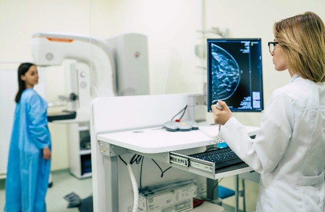
Better imaging for breast cancer diagnosis and treatment
TRI-based researchers from QUT are refining portable imaging technology to create 3D images of breast tissue, with the aim of improving cancer diagnosis and treatment.
QUT’s Professor Rik Thompson and Dr Konstantin Momot are collaborating with CSIRO’s Associate Professor Jason Dowling and Dr Hollie Min, and Metro South Health’s Dr Thomas Lloyd, with TRI and Australian e-Health Research Centre funding.
Key points
- QUT researchers based at TRI are part of a collaboration developing accurate, low-cost medical imaging technology to map breast tissue in 3D.
- The imaging technology has the potential to identify the level of breast density, monitor prevention strategies and track the efficacy of treatments.
- The technology has no ionising radiation, meaning it should be safe and non-invasive.
- Because the technology is portable, it has the potential to be used outside of hospitals, in mobile clinics and in rural and remote communities.
The problem with dense breast tissue
More than 20,000 people were diagnosed with breast cancer in Australia in 2022, making it among the most commonly diagnosed cancers. More than 3,000 died of breast cancer in the same year.
Breast screening is among the most successful techniques for early detection, leading to better treatment options and improved survival rates.
But almost half of all women are in the higher categories of mammographic breast density, which is a known risk factor for breast cancer.
The dense breast tissue appears white on a mammogram, as do breast cancers, making the images more difficult to interpret and increasing the risk of a cancer going undiagnosed. Yet mammography remains the best screening method for breast cancer, even for women with dense breasts.
Professor Thompson says women with increased risk factors for breast cancer, including high breast density, can benefit from supplementary screening, such as clinical MRI and ultrasound.
“It’s not dense to touch, it’s not a lump and it’s not firm – but it absorbs x-rays and shows up as white,” he says.
Embracing imaging in 3D
Professor Thompson is leading a research team aiming to develop imaging technology that will enable breast tissue to be mapped in 3D.
“Magnetic resonance imaging (MRI) is capable of mapping the 3D distribution of mammographic density within the breast as well as identifying benign and malignant abnormalities,” he says.
MRI does not use ionising radiation, a type of energy released by atoms which can increase the risk of longer-term effects such as cancer, if used frequently.
However, cost is a major impediment to the use of MRI for routine mammographic density screenings.
Professor Thompson says his research group has pioneered biomedical applications of portable NMR as a viable alternative for breast tissue imaging.
“It is a low-cost, mobile technology based on the same fundamental physics as conventional MRI.
“Our results demonstrate the promise of the imaging technology for accurate, safe and low-cost quantitative assessment of mammographic density.”
Advancing research from 3D models to actual patients
Professor Thompson’s team is using 3D-printed scaffolds and water-based gels to create models that emulate the 3D distribution of dense tissue, called breast density phantoms.
They are imaging the phantoms at TRI to assess their density, sharing analysis and generated data with CSIRO collaborators, and taking what they learn to develop methods for imaging human tissue.
The research involves collaborators at Princess Alexandra Hospital to ensure methods developed will be applicable for imaging in patients undergoing cancer preventative therapy or anticancer adjuvant hormonal therapy.
Existing clinical collaborations are leveraged for the project, including with surgeons, radiologists and pathologists. Further collaborations between Professor Thompson and clinicians in Perth are assessing whether change in mammographic breast density may also indicate that other types of therapy are working.
Supporting the work is $100,000, jointly awarded by TRI and the Australian e-Health Research Centre at CSIRO, as part of funding for projects aimed at solving a healthcare challenge. The grants support research teams with a TRI-based researcher, CSIRO scientist and a TRI partner clinician from Metro South Health.
It builds on earlier $300,000 in support from the Princess Alexandra Hospital Research Foundation and a $90,000 TRI Spore Grant to develop the technology.
The research has resulted in a new collaborative project to identify the molecular and cellular basis of mammographic brightness and Dr Sandy Minck’s recruitment as a consumer advocate for both projects.
Healthcare application beyond breast cancer
Beyond application in breast cancer, the research aims to develop portable imaging techniques suitable for use in diagnosing and monitoring a variety of other diseases.
“The approaches developed in this project have the potential to pave the way for low-cost portable NMR instrumentation to be used for a broad range of quantitative medical imaging,” Professor Thompson says.
“It has the potential to expand the availability of medical imaging in remote and rural locations, in primary emergency care, and for routine low-cost screening.”
Dr Momot is also investigating the use of the portable NMR for assessment of tendons, and it potentially has other applications in skin cancer assessment and monitoring.
“There are quite a few other prospective applications of Portable NMR, including monitoring of hydration or blood oxygenation levels, detection of internal haemorrhage in trauma patients, as well as liver or brain imaging,” Dr Momot says.
“Some of these applications require custom-built portable NMR instrumentation, but the technique itself has very broad applicability.”
Publications
Foongkajornkiat S, Sokolowski KA, Stephenson J, Lloyd T, Hugo HJ, Thompson R, Momot K. (2024). Quantitative measurement of mammographic density in breast-tissue explants using portable NMR: Precision and accuracy. Magnetic Resonance in Medicine, 92(1), 374–388. DOI: https://doi.org/10.1002/mrm.30040
Faheem M, Tam HZ, Nougom M, Suaris T, Jahan N, Lloyd T, Johnson L, Aggarwal S, Ullah MZ, Thompson EW, Brentnall AR. (2024). Role of Supplemental Breast MRI in Screening Women with Mammographically Dense Breasts: A Systematic Review and Meta-analysis. Journal of Breast Imaging, Article wbae019. DOI: https://doi.org/10.1093/jbi/wbae060
McKay-Parry ND, Blick T, Foongkajornkiat S, Lloyd T, Thompson EW, Hugo HJ, Momot KI. (2022). Portable NMR for quantification of breast density in vivo: Proof-of-concept measurements and comparison with quantitative Magnetic Resonance Imaging, 92, 212–223. DOI: https://doi.org/10.1016/j.mri.2022.07.004