Expertise
Mark Scott
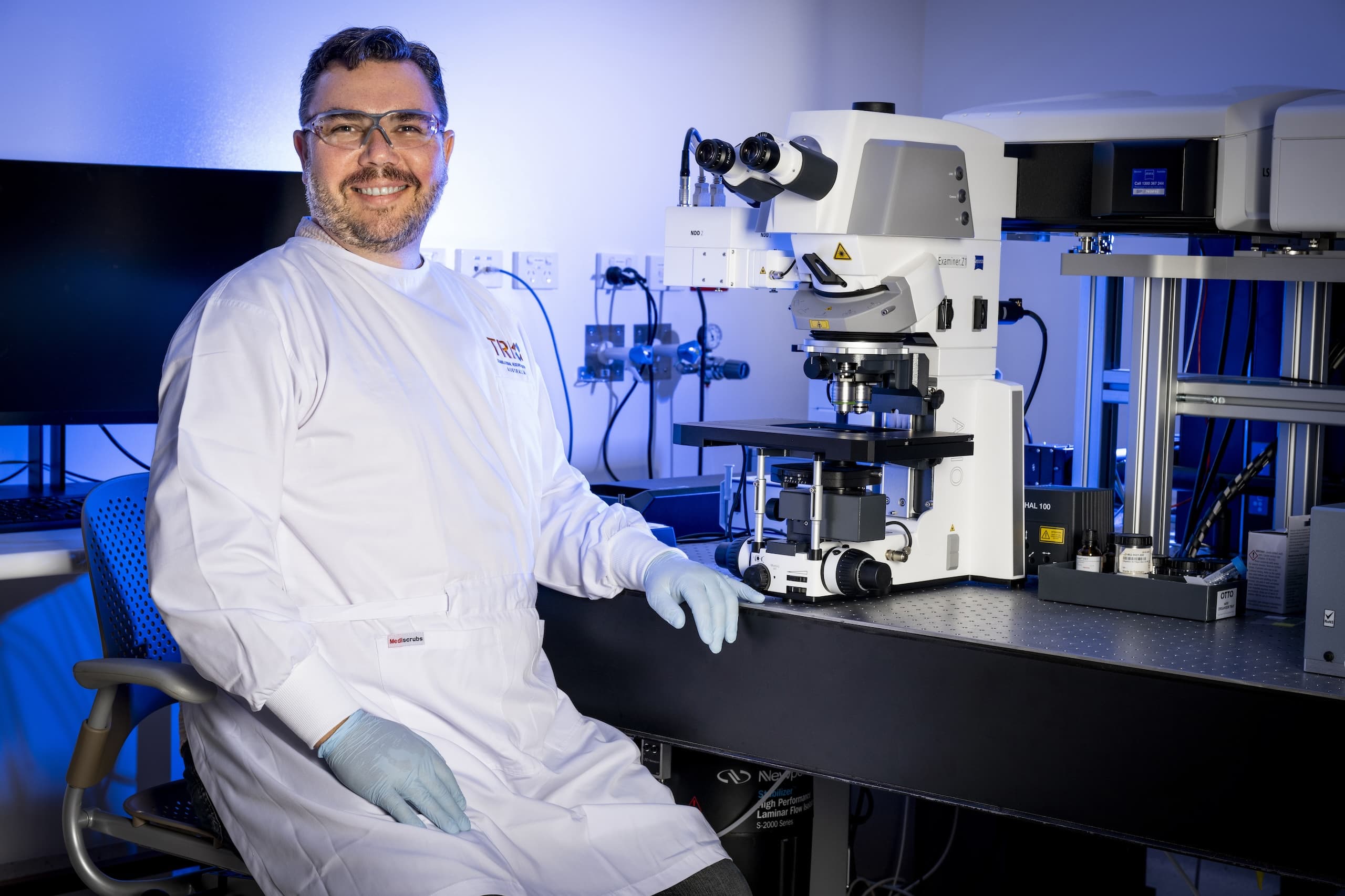
Senior Microscopy Scientist
Experience
Mark started his career in the electron microscopy department of QEII Medical Hospital at UWA before moving to the UK and working on the NIH Human Brain Project in Newcastle-upon-Tyne, mapping 3d gene expression data of the developing human brain. He has since worked in several research institutes around Australia and the UK, including Imperial College London, WEHI in Melbourne and the IMB at UQ and has been instrumental in establishing intra-vital two-photon microscopy techniques at a number of these institutions and published papers on HSC imaging in bone marrow.
Mark has also developed a solid understanding of cleared tissue imaging and clearing methods, as well as super-resolution techniques. He also has several years of developing image analysis pipelines and automated scripts for both his own projects and those of researchers he has helped in core facility environments.
Meet the Team
The TRI Microscopy team are able to facilitate a wide range of research opportunities within the core facility. The team’s collective experience has been developed through academic research positions as well as working within a number of different core facilities. Staff also have a significant amount of complimentary experience in areas such as electron microscopy, flow cytometry, histology and cell culture, allowing them to provide holistic experimental support and facilitate inter-disciplinary collaborations. Facility staff are distinguished by several high-profile publications in microscopy applications and are eager to help researchers with pushing the boundaries of their work.
The facility staff are also well versed in a wide range of research techniques and are able to offer assistance with experimental design and troubleshooting, facility inductions and system training, sample preparation and tissue clearing and image analysis software including machine learning segmentation tools. The facility also offers bright-field and fluorescence slide scanning as a fee for service.
Cameron Flegg – Microscopy Officer: Cameron was first exposed to light microscopy at undergraduate and honours level, completing courses in fluorescence and electron microscopy and an honours project specifically in microscopy techniques, looking at host-pathogen interactions. Completing his PhD in host-pathogen interactions at the IMB (macrophage-bacteria) he utilised extensive microscopy in confocal, electron and live imaging microscopy. Further research experience was gained in host-pathogen interactions, cell trafficking, nuclear-cytoplasmic regulation of proteins in cancer and bone biology/regulation of osteoclasts. All projects have been microscopy intensive in confocal, widefield, live imaging and electron microscopy. He has also previously managed Imaging and Flow facilities and now has over 20 years’ experience in imaging techniques and continues to expand his knowledge and understanding of image analysis tools.
Andy Wu – Core Facilities Officer: Andy gained his PhD and worked as a Postdoctoral Researcher on Osteoimmunology and Cancer Metastasis using flow cytometry and immunohistochemistry techniques to analyze pre-clinical mouse samples. He has gained experience using wide-field and confocal microscopes to examine protein expression on histological samples. Andy has worked across several core facilities within TRI, including the Histology core and continues to share his time between Flow Cytometry and Microscopy cores here building experience in use training on several wide field systems, Incucytes and slide scanning as well as instrument troubleshooting.