Login
Contact

Explore

Explore

About
  • People and Partners
  • TRI Building
  • Reconciliation Action Plan
  • Strategic Plan
  • TRI Foundation
  • Careers
Research
  • Collaborate
  • Discovery
  • Translate
  • Engage
  • Research Areas
Clinical
  • Translational Trials
  • Clinical Research Facility
  • TRI at Children’s
Industry
  • TRI Incubator
  • The Precinct
  • Residents
Manufacture
  • New cGMP Facility
Facilities
  • Cleanroom
  • Flow Cytometry
  • Histology
  • Microscopy
  • Proteomics
  • Preclinical Imaging
  • Biological Research
  • Gnotobiotics
  • Human Immune Model
Get Involved
  • Health Consumers
  • SPARQ-ed
Resources
  • News
  • Events
  • Case Studies
  • Grants and Funding
  • Education and Training
+61 7 3443 7000info@tri.edu.au37 Kent Street, Woolloongabba QLD 4102 Australia

Acknowledgement of Country

TRI acknowledges the traditional custodians of the land we are on today, the Turrbal and Jagera people and we recognise their continuing connection to land, waters and community. We pay deep respect to them and their cultures; and to Elders past, present and emerging.

© Translational Research Institute Pty Ltd atf the Translational Research Institute Trust|ABN: 58 155 991 662
Privacy policy|Terms of Use|Contact Us
Back to equipment

BX63 Upright Epifluorescence

Olympus BX63 Upright Epifluorescence

Olympus BX63 Upright Epifluorescence

Fully motorised brightfield and fluorescence scanning of tissue sections or cells on slides.

An advanced imaging tool ideal for a broad range of imaging applications including time-lapse movies, multiwell plate scanning, image stitching and tiling, and cell counting.
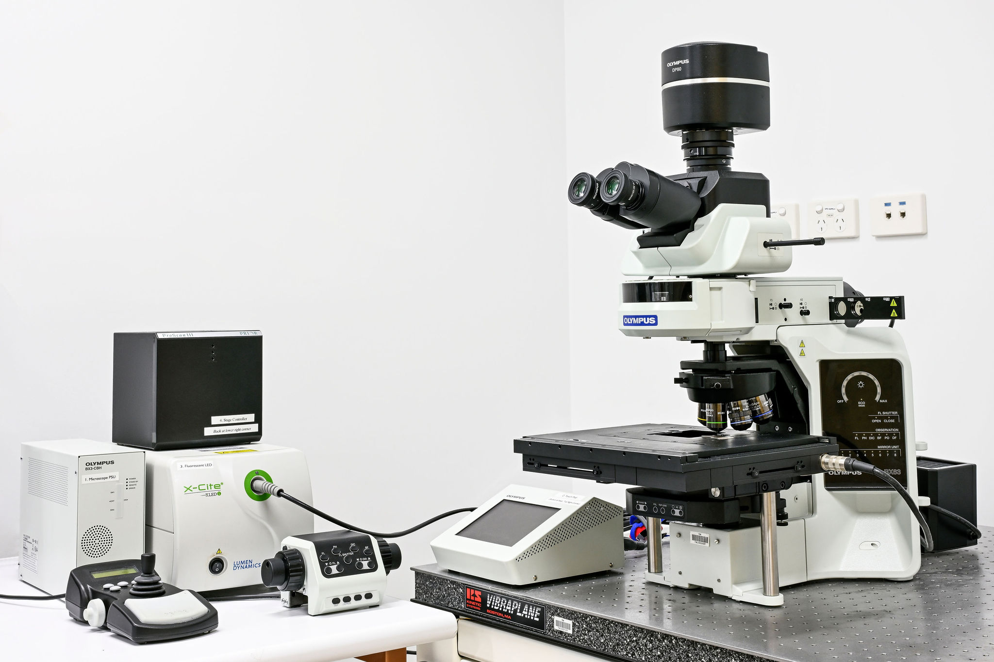
 

Features

  • Objectives: 4X, 10X, 20X and 40X, 60X Oil and 100X Oil
  • Camera/Detector: DP80 monochrome/colour camera
  • Light Source: Halogen lamp for brightfield. X-Cite LED for fluoresence (DAPI, GFP, CY3, CY5)
  • Software: Cell Sense
  • Application: Slides only, automated imaging of multiple colours and single slide tile scanning

Training

Microscopy instruments can only be booked once training and certification has been completed. Contact the TRI Microscopy team for further information.

Need more help?

For more information on instruments, services and facility charges contact the Microscopy Facility Manager.