Molecubes
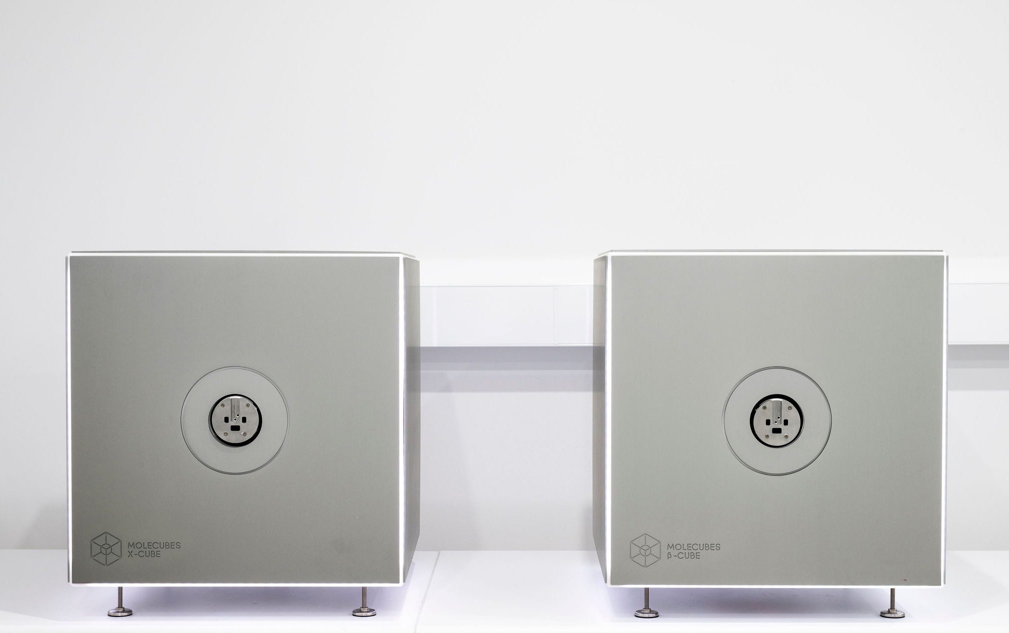
Molecubes β-Cube and X-Cube (µPET-CT)
Molecubes β-Cube and X-Cube (µPET-CT)
The benchtop Molecubes µPET-CT system co-registers sequentially acquired PET and CT data from anaesthetised small laboratory animals.
The PET system (β-Cube) provides sub-millimeter spatial resolution, with a maximal Field of View (FOV) of 130mm (axial) x 72mm (transaxial).
β-Cube Features
High-throughput imaging is achievable due to high detector sensitivity (short acquisition times), simultaneous imaging capability, and a user-friendly software graphical interface. Our facility is licensed to handle multiple PET radioisotopes including F18, Zr89, Ga68 and Cu64. Common PET-CT applications include glucose uptake, tumour-specific imaging, bio-distribution of newly developed radiotracers, and brain dopamine metabolism.
X-Cube Features
Utilises a variable X-ray source (20-80kV) enabling rapid whole animal imaging through a large FOV of 63mm (transaxial) x 200mm (axial). CT images are typically reconstructed at 50µm voxel size resolution. The novel respiration gating functionality minimises breathing artefacts, providing clear CT images. The X-Cube can be used independently, and common CT applications include longitudinal assessment of osteogenesis, calcification of biomaterials, osteoporosis, patient-derived explants (samples inside plates), and healing in bone defect models.